Category Archives: Orthopedics & Neurological Rehabilitation

What a Pain in the Neck

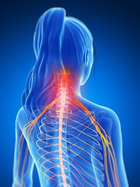
Neck pain is a common reason for doctor visits. It is so common that the topic made it to the Washington Post recently in an article titled “What you need to know about that pain in your neck.”

Here are some facts about neck pain:

  • A common cause of neck pain is poor posture during work, rest or leisure.
  • Lifting or carrying heavy objects can strain muscles connecting your arm/shoulder and neck.
  • Even light weight (or un-weighted) repetitive motions can cause neck pain.
  • Excessive tension or stress frequently causes us to hold our jaw, neck and shoulders in a way that leads to pain and headaches.
  • The joints of the neck allow a great deal of motion. That is why necks are vulnerable to injury with trauma.

What you need to know before you go to the doctor:

  • Majority of neck pain episodes resolve in 2-3 weeks without imaging studies.
  • The older we are the more likely neck pathology will be seen on imaging studies even on those people without neck pain.
  • Over the counter analgesics (Acetaminophen, Ibuprofen, Naproxen Sodium) are more effective in treating neck pain than muscle relaxants.
  • Sleeping with too many pillows or a pillow that is too firm can cause neck pain.

What you can do:

  • Use heat or cold pack to soothe muscle aches. Use the type that feels best to you.
  • Decrease unnecessary weight in your handbag, book-bag or briefcase.
  • Remind yourself to relax your neck, jaw and shoulder muscles while trying to maintain good posture.
  • Consider Physical Therapy to help improve muscle and joint balance via exercises for your neck and shoulder girdle.
  • If pain has not resolved in time, or if your pain was caused by trauma, see your physician or urgent care clinic.


Written by: Miriam Graham, PT, DPT
Edited by: Claire Agrawal, PT, DPT

Photo Credit:
http://media2.mindandbodychiro.com.au/wp-content/uploads/2015/01/img-neck-pain-spreading-shoulders-450×600.jpg

Link to Washington Post article:
https://www.washingtonpost.com/national/health-science/what-you-need-to-know-about-that-pain-in-your-neck/2016/04/11/5666e97c-a2a3-11e5-b53d-972e2751f433_story.html

New ACL Surgery May Improve the Lives of Many

ACL Knee Surgery Picture Blog, May, 2016
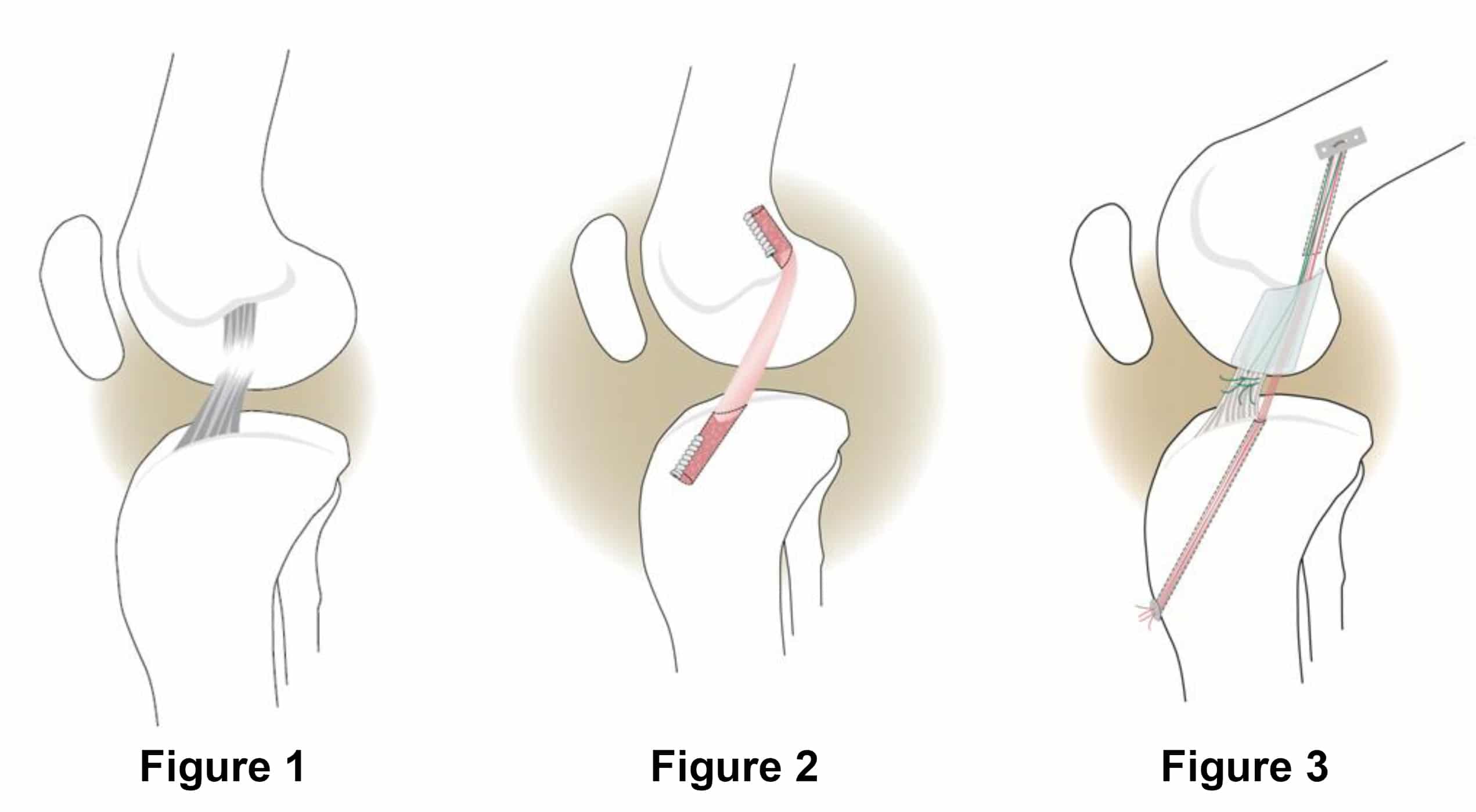
The anterior cruciate ligament (ACL) of the knee is one of four ligaments between the femur and the tibia that helps keep the bones together and stable during movement. Injury to this ligament can lead to knee pain, a feeling of your knee buckling or giving out from under you, and decreased knee range of motion. Spraining or tearing of the ACL is one of the most common knee injuries in humans. Depending on the severity of the injury and the person’s age, physical therapy may be the first treatment option to stabilize the knee. If symptoms do not improve with conservative management, surgery is often considered next.

Currently, the standard surgical technique is called ACL reconstruction and involves removal of the torn ends of the ligament and replacement with a tissue graft. A tissue graft is tissue from another part of the body (usually hamstring tendons) or a cadaver. The tissue graft acts as scaffolding for a new ligament to grown on. Recovery is lengthy and patients are at a higher risk of developing arthritis 15-20 years post surgery (Boston Children’s Hospital).

A new surgery, called “bridge-enhanced ACL repair, or BEAR,” has been developed by Dr. Martha Murray and her team at Boston Children’s Hospital. The technique places stitches and a sponge injected with the patient’s own blood between the ends of the torn ACL to stimulate healing of the original ligament instead of replacing it. Over six-to-eight weeks, the ends of the torn ACL grow into the sponge and reconnect.

There is potential for the BEAR technique to allow quicker recovery of strength after ACL surgery and possibly reduce the long-term risk of arthritis in the injured knee. At this time the surgery is experimental and being performed through clinical trails to determine long term risks and benefits. To learn more about the clinical trials, click here.


Written by: Kristen Bissell
Edited by: Claire Agrawal, PT, DPT

Clinical Trial link:
http://www.childrenshospital.org/centers-and-services/anterior-cruciate-ligament-program/bridge-enhanced-acl-repair-trial

Photo Credit:
http://www.childrenshospital.org/~/media/centers-and-services/departments-and-divisions/sports-medicine-division/bearimage1.ashx?la=en

What is Diastasis Rectus Abdominis?

Most people know that the abdominal muscles can be toned and displayed as a “six-pack.”  Fewer people know that the same muscles can also separate from each-other, right down the midline, forming a diastasis rectus abdominis (DRA). diastasis-recti

Diastasis means separation. Rectus abdominis refers to muscles that make up part of the abdominal layer closest to the skin. They are the vertically oriented muscles in the picture above, comprised of a right and left side. The two strips of muscle are connected to each-other by connective tissue (fascia). In the formation of a diastasis rectus abdominis, the connective tissue stretches and the muscles elongate.

Q: What are the Risks Associated with Having a Diastasis Rectus Abdominis?

A: Increased risk of injury to your back from decreased core support.

Muscles function best at optimal length. If the rectus abdominis muscles are asked to work in an over-stretched position as in a DRA, they will not contract as efficiently. The result is less core support of your spine and pelvis. Decreased core strength or core muscle imbalance is associated with risk of injury, especially to your back.

In addition to the risk of injury, some people simply dislike the appearance of a protruding abdomen.

How can a Diastasis Rectus Abdominis Form?

  • Improper lifting techniques and chronic straining (with lifting or to move bowels)
  • Pregnancy
  • Significant weight gain
  • Weakened abdominal muscles or connective tissue disruption from surgery
  • Idiopathic, born with it

What can be done about a Diastasis Rectus Abdominis?

Treatment options include physical therapy, wearing an abdominal brace, or surgery.

Physical therapy treatment for DRA may include the following:

  • Identification of movements and postures that exacerbate the DRA and education on how to avoid or modify these factors. This includes education on proper lifting/carrying/lowering body-mechanics.
  • Exercise prescription focused on helping the rectus abdominis muscles activate as well as balancing the strength and coordination of deep core muscles (e.g. pelvic floor muscles, transverse abdominis, multifidi, and diaphragm).
  • Resolution of myofascial trigger points in the core musculature
  • Abdominal taping or bracing to bring the rectus muscles back aid optimal length for function

Appropriate treatment will depend on the severity of the separation and how long it has been that way.                           Consult your medical team to determine if you have a diastasis rectus abdominis and what is the best approach to treat it.


Written by: Claire Agrawal, PT, DPT


Photo credits: Diastasis Rectus Abdominis: http://www.applehealthwellness.com/wp-content/uploads/2013/11/diastasis-recti.jpg

Ehlers-Danlos Syndrome (EDS) – Pain Management with Your Physical Therapist

postone

*    Ehlers-Danlos Syndrome, or EDS is a hereditary disorder affecting the body’s ability to produce collagen. Collagen is the most abundant structure in the body and is a component of bone, cartilage, tendons, ligaments, and organs.
*    There are six types of EDS. The three most common types are:
o Hypermobility type of EDS
o Classic type of EDS
o Vascular type of EDS
*    Signs and Symptoms vary by type and may include: hyper-extensible joints, muscles, and skin; chronic joint pain; easy bruising; chronic fatigue; gastrointestinal mobility disturbances; and autonomic dysfunction (such as hypotension and difficulty with body temperature regulation).
*    It is important to see your doctor for a diagnosis of EDS, as man other conditions share the same signs and symptoms.
*    Once diagnosed, you may be referred to a team of health professionals besides your primary doctor. This team may include a sleep specialist and a physical therapist.
*    People with EDS may avoid physical activity due to pain associated with previous trials of exercises. Doing so, however, leads to muscle weakness and further exacerbates joint instability and pain. Your physical therapist can help you with pain management, finding safe ranges to work your muscles, and fun ways to stay active.

Fall Prevention Tips – Balance Exercises and Suggestions from your Physical Therapist

post2 - holding hands

Every other Wednesday, a group of senior citizens gather at the Shri Mangal Mandir Hindu temple to learn about a specific health topic.

o Human balance is a complex system, depending on vision, inner ear or vestibular function, information from muscle and joint receptors, muscle strength, and the ability to integrate information from all these inputs to tell the brain where the body is in space.
o Balance exercises are as important to the body as cardiovascular and strength training. They can be incorporated into your weekly exercise routine as you age to help prevent falls.
o Some ways to incorporate balance training include walking on uneven surfaces, doing Yoga or Tai Chi, or just keeping your balance as you close your eyes or turn your head. ***Not all balance exercises are safe for everyone, so speak with a physical therapist or other health provider if you have a history of falls or dizziness before trying these suggestions.
o You can reduce your risk of falls in your home by:
* Making sure you have good lighting and handrails on your stairwells both indoors and outdoors
* Putting in night lights along the path from your bedroom to your bathroom
* Moving objects you use frequently to a more accessible location such as a lower shelf in your cupboard to avoid going on tiptoes or bending down low
* Removing throw rugs or other slippery objects from the floor and installing grab bars in the bathroom
For more information about balance and fall prevention, the CDC website is a valuable resource. Click here for an overview of falls in older adults. Link for the overview of falls in older adults:

http://www.cdc.gov/HomeandRecreationalSafety/Falls/adultfalls.html